Infographic: How a Glutamate Sensor Tracks Synapses

A third generation glutamate sensor with a fluorescent readout offers insights into neuronal communication.

Ultrafast glutamate sensors resolve synaptic short-term plasticity

Glutamate signalling in non-neuronal tissues: Trends in Pharmacological Sciences

Figure 1, Schematic drawing of a glutamatergic synapse, with postsynaptic AMPA, NMDA, KA and metabotropic receptors - Jasper's Basic Mechanisms of the Epilepsies - NCBI Bookshelf
Glutamate sensing in biofluids: recent advances and research challenges of electrochemical sensors - Analyst (RSC Publishing) DOI:10.1039/C9AN01609K

Schematic representation of a glutamatergic neuronal synapse, showing

Infographic: How a Glutamate Sensor Tracks Synapses

Electrochemical Biosensors for Whole Blood Analysis: Recent Progress, Challenges, and Future Perspectives
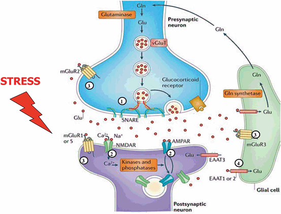
Synaptic Stress, Changes in Glutamate Transmission and Circuitry, and Psychopathology

Detection of glutamate release from neurons by genetically encoded surface-displayed FRET nanosensors
Infographics Articles The Scientist Magazine®
IJMS, Free Full-Text

Single synapse glutamate imaging reveals multiple levels of release mode regulation in mammalian synapses - ScienceDirect

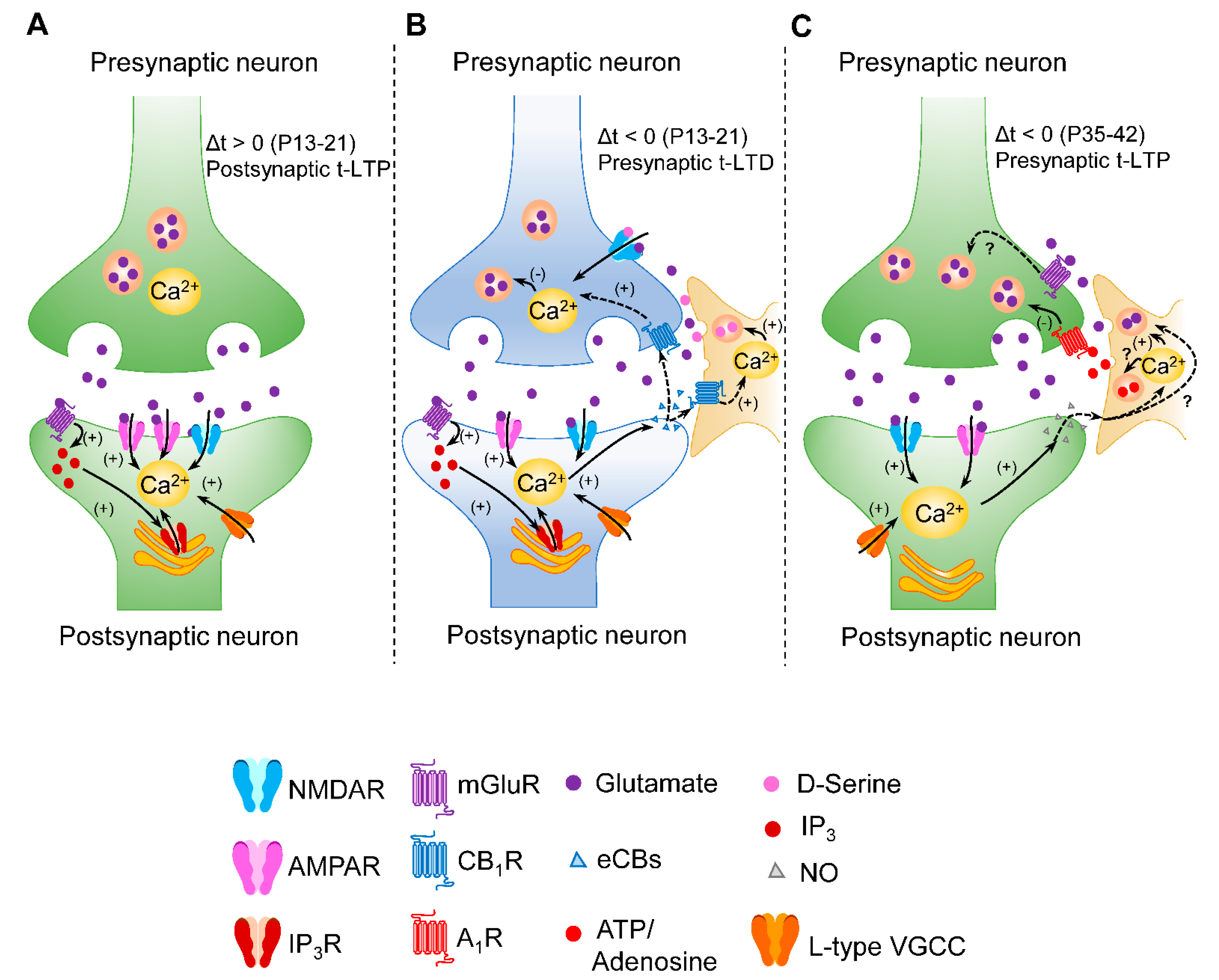
Infographic: Reverse Signaling Between Neurons